Diabetes mellitus is among other common chronic diseases affecting people’s health. Despite adults are the most diagnosed age group, diabetes can actually develop in anyone at any age, depending on the types of diabetes: type 1 and type 2 diabetes. Type 2 diabetes is an impairment of using insulin to regulate and use sugar (insulin resistance). As the most common type of diabetes, it is often found in people aged over 40. Whereas type 1 diabetes or insulin-dependent diabetes usually occurs in adolescents and children aged younger than 10 when the pancreas produces insufficient or no insulin. Nevertheless, interesting findings derived from clinical studies reveal that the incidence of type 2 diabetes has increased among obese children. The main contributing factors are related to unhealthy lifestyles, including overconsumption of carbohydrate and sugar, lack of exercise and sedentary behaviors, such as sitting or lying down while engaged in watching television, playing video games and using a mobile phone or computer for much of the day.
Diabetes and eyes
Diabetes is defined as a metabolic disease present with an elevated blood sugar level. In normal conditions, insulin as a crucial hormone works to lower the amount of sugar in the blood circulation by moving sugar from the blood into the cells to be stored or used for energy. With diabetes, the body either does not produce sufficient amount of insulin (type 1 diabetes) or cannot use insulin effectively due to insulin resistance (type 2 diabetes). Instead of being transported into the cells, sugar builds up in the bloodstream, causing high blood sugar levels. Overtime, regardless of the type of diabetes, uncontrolled blood sugar can potentially lead to blood vessel damages, causing a wide range of diabetes complications affecting different organs, such as the kidney, heart and nervous system as well as the eyes. Eye damages caused by diabetes (diabetic eye diseases) can interfere with several parts of the eyes, including the muscle of the eye, lens, optic disc, optic nerve and retina.
Diabetes and blindness
Diabetes is recognized as one of the leading causes of preventable blindness in adults age 20–74. Over time, high blood sugar levels from diabetes lead to damage of the retina, resulting in vision loss. People with diabetes are 25 times more likely to experience blindness than people without diabetes.
Diabetes retinopathy
Diabetic retinopathy is a complication of diabetes caused due to prolonged high blood sugar levels damaging the back of the eye or retina, an essential part of the eye that enables vision. There are 2 main conditions that lead to an impaired vision. However, these conditions are independent of another.
- Macular edema
During early stage of diabetic retinopathy, the walls of blood vessels in the retina are weakened. Tiny bulges in the blood vessels protrude from the walls of the smaller vessels and may leak fluid and blood into the retina. This leakage can cause swelling of the macula. In some cases, retinal blood vessel damages might lead to a buildup of fluid or edema in the macula. If macular edema develops, it often distorts central vision. - Proliferative diabetic retinopathy (PDR)
As an advanced form of diabetic retinopathy, damaged blood vessels become completely occluded, causing insufficient retinal blood supply and inducing the growth of abnormal vessels in the retina. These newly formed blood vessels are fragile and susceptible to leak into vitreous –the jellylike substance that fills the center of the eye. Eventually, scar tissue, as the result of the growth of these new blood vessels, can cause the retina to detach from the back of the eye. Retinal detachment affects vision by causing blurred vision or darkness or curtain-like images in the vision. If not treated, it can lead to blindness.
For more information, please click: Do Not Overlook Diabetic Retinopathy As It Can Lead To Complete Vision Loss!

Neovascularization of the iris
Neovascularization of the iris defined as blood vessel proliferation along the surface of the iris is a complication caused due to diabetic retinopathy. Iris neovascularization can be induced by lack of adequate blood supply and oxygen (hypoxia). As a result, small blood vessels develop on the anterior surface of the iris in response to retinal ischemia. These newly formed blood vessels may occlude the drainage of aqueous humor, leading to a sudden rise of an intraocular pressure which is known as neovascular glaucoma. Neovascular glaucoma is generally associated with poor visual prognosis and less response to medications, compared to other types of glaucoma. Diabetic retinopathy, which is the leading cause, must be treated.
Cataracts
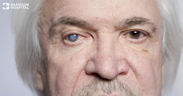
Cataracts, the clouding of the lens of the eye, can potentially impair vision.
The lenses within the eyes are clear structures that provide sharp vision, but they tend to become naturally cloudy as we age. Hinging on duration of diabetes and ability to control blood sugar levels, people with diabetes can develop cataracts at an earlier age than non-diabetic people. When prescription glasses cannot clear the vision, the only effective treatment for cataracts is surgery. Cataract surgery is often considered when it begins to affect quality of life or interfere with ability to perform normal daily activities, such as reading or driving at night.
Diabetic keratopathy
In comparison to people without diabetes, diabetic patients are more likely to have corneal injuries or ulcers easily due to diabetic corneal alterations, including delayed epithelial wound healing, recurrent erosions, loss of sensitivity, and tear film changes. As a result, it typically takes longer time to complete corneal healing process while the risk of corneal infections also rises because of delayed healing.
Paresis of extraocular muscle
Extraocular muscle paresis develops when extraocular muscle is weak due to lack of muscle function. The damage of blood vessels caused by diabetes usually involves the third (oculomotor), fourth (trochlear) and sixth (abducens) cranial nerves which control the movement of the eye. Double vision is the main characteristic symptom. Paresis of extraocular muscle is usually present with good prognosis in which reversible symptom typically subsides within 3 months. To temporarily correct double vision, wearing prism glasses can be considered. If double vision is persistent longer than 6-12 months without any improving sign, surgery to repair extraocular muscles might be an alternative.
Preventing diabetic retinopathy
- Keep blood sugar, blood pressure and blood lipids under control
- Regularly exercise
- Refrain from smoking and alcohol
- Pay extra attention to vision changes, especially when vision suddenly becomes unclear, spotty or hazy.
- Schedule a yearly eye exam despite no abnormal sign has yet exhibited.
Diabetic patients need to undergo an annual eye examination with pupil dilation in order to screen for diabetic retinopathy. During this eye exam, the ophthalmologist begins to examine the eye and eye drops are placed in the eyes to dilate the pupils, allowing the ophthalmologist a better view the inside of the eyes. The eye drops used to dilate the pupils can cause blurred vision until their pupil dilating effects wear off, taking approximately 4-6 hours. Hence, patients are not advised to drive themselves after having their eyes dilated and caregivers or relatives should accompany the patients after the test. Without any ocular abnormality, only a yearly comprehensive eye exam is required. In case that diabetic retinopathy is detected, treatment and follow-up schedules will be arranged, based upon its severity.


