Gallstones or Cholelithiasis
- Risk factors: Gallstones are hardened deposits of digestive fluid mainly made of cholesterol which is formed in the gallbladder. This condition has been commonly found in women above 40. Particular risk factors include hypercholesterolemia (high level of blood cholesterol), being diabetic, taking medications that contain estrogen such as oral contraceptives or hormone replacement therapy, having had several children and drug-induced gallstones such as some lipid lowering agents.
- Signs and symptoms: Gallstones sometimes do not manifest any specific symptoms. Mild to moderate symptoms start from abdominal discomfort to flatulence and pain after consuming high-fat foods. A lot of people often mistake gallstones for peptic ulcers since both conditions have similar symptoms.
- Treatment: To diagnose gallstones, an upper abdominal ultrasound is highly recommended in order to clearly visualize the stones that have formed in the bladder. Treatment for gallstones is to surgically remove the gallbladder called “cholecystectomy”. Laparoscopic cholecystectomy is a minimally invasive surgery to remove gallstones through smaller incisions, compared with an open surgery which a large cut is required. Since gallstones frequently recur, once gallbladder is removed, bile flows directly from the liver into the small intestine, rather than being stored in the gallbladder, therefore it helps to reduce risks of serious complication caused by gallstones that might lodge in a bile duct and induce a blockage, eventually results in bile duct inflammation and infection, pancreatitis or cholecystitis (an inflammation of gallbladder).
Acute Cholecystitis
- Risk factors: chronic abdominal pain or untreated gallstones
- Signs and symptoms: Aggravating symptoms include severe and sudden pain in the upper right abdomen especially when breathing in and out, back pain between shoulder blades, high fever with chills, jaundice with a yellow tinge to the skin and whites of the eyes, pale stool, nausea, vomiting and colic pain that radiates to the whole abdomen.
- Treatment: There are 2 main surgical techniques to remove gallbladder: open cholecystectomy and laparoscopic cholecystectomy. Laparoscopic cholecystectomy is a minimally invasive surgery to remove gallbladder which is performed by highly experienced and well-trained surgical team. Through smaller incisions, the procedure involves laparoscope, a narrow tube with a camera, 3D and 4K Ultra HD technology that significantly enhance the surgical accuracy and safety, resulting in satisfactory outcomes with minimized chances of infection caused by gallbladder ruptures.
- Outcome measurement of laparoscopic cholecystectomy
- 100% Patient’s ability to walk within 4-6 hours after operation in healthy candidates for surgery.
- 93% Success rate of operation incluing in acute cholecystitis
- 0% Major injury of bile duct.
*Reference: Statistical data obtained from Surgery Center, Bangkok Hospital 2018.
Incarcerated Hernias
- Causes: Hernias occur when an organ especially small intestine protrudes through a weakened spot or tear in the abdominal wall. Hernias are commonly caused by a combination of muscle weakness and increased abdominal pressure. Hernias cause a bulge or lump in the affected area such as groin, diaphragm or surgical incision which is not properly closed.
- High risk group: Groin hernias (inguinal hernia) is commonly found in middle age or elderly men with risk factors including lifting heavy objects, being obese, being diagnosed with benign prostatic hyperplasia (BPH), chronic respiratory diseases such as emphysema and chronic bronchitis and chronic constipation that causes the strain during defecation and increases an abdominal pressure. As a result, the muscles of abdominal wall become weakened and a part of small intestine protrudes through this weak area, presented as a bulge or lump. An incompletely healed surgical wound in the abdomen can also cause incisional hernia. Hernias often found in women include femoral hernias, hiatal hernia (diaphragm hernias) and obturator hernia (hernia of the pelvic floor). In addition, umbilical hernia in babies can develop when a portion of the lining of the abdomen, part of the intestine or fluid from the abdomen comes through the muscle of the abdominal wall, presented as an abnormal bulge that can be seen or felt at the umbilicus (belly button).
- Signs and symptoms: The most common symptoms are pain or discomfort (usually at lower abdomen), weakness or heaviness in the abdomen, burning or aching sensation at the bulge. Hernias can be particularly felt during standing up, bending down or coughing. Hernias typically flatten or disappear when they are pushed gently back into place or when patients lie down. If the protruding intestine is not pushed back in place, the contents of hernia might be trapped in the abdominal wall, then becoming strangulated which cuts off blood supply to surrounding tissue that is trapped, leading serious complications. If protruding bulge cannot be pushed back, immediate medical attention must be sought in order to receive accurate diagnosis.
- Treatment: Differential diagnosis must be made in order to distinguish other diseases which might have overlapping symptoms such as tumors, swollen lymph nodes, testicular torsion or spermatic cord. An incarcerated hernia occurs when herniated tissue becomes trapped in the weak point in the abdominal wall and cannot easily be moved back into place, As a result, it can obstruct the bowel, leading to intestinal obstruction. The goal of treatment primarily aims to push back protruding intestine or tissue into place while preventing recurrences. If the protruding part cannot be pushed back, incarcerated intestine must be surgically removed by using “laparoscopic totally extraperitoneal hernia repair or TEP”. During this minimally invasive procedure, instead of making an open cut, the surgeon operates through 3 small incisions in the abdomen. A small tube attached with a tiny camera (laparoscope) is inserted into one incision. Guided by this camera, the surgeon then inserts tiny instruments through other incisions to repair the hernia by pushing protruding tissue back in place. To enhance the strength of muscles in the abdominal wall, during this surgery, synthetic mesh, sized 10×15 cm. will be implanted to provide additional support to weakened areas. Not only reducing the tenderness and pain after surgery, but mesh repair done by highly experienced surgeons also significantly helps minimizing the chances of hernia recurrence. Due to the advancements in laparoscopic instrument with 4K Ultra High Definition, it enables surgeons to clearly visualize the surgical field in the abdominal cavity including internal organs, blood vessels and nerves. As a consequence, it helps enhancing surgical accuracy, resulting in smaller incisions, less pain, less blood loss and reduced post-operative complications as well as a faster recovery time and a quicker return to normal activities.
Liver, pancreatic and biliary diseases
- Acute hepatitis: Acute viral hepatitis is inflammation of the liver caused by viral infection, fatty liverm side effects of certain drugs, the ingestion of toxic substances and long-term effects of alcohol consumption. Acute hepatitis results in enlarged liver, jaundince with yellow discoloration of a skin and eyes, weight loss and pain in the upper right part of the abdomen. If left untreated, it tends to progress to chronic hepatitis which permanently damages the liver tissues, causing cirrhosis, a late stage of scarring (fibrosis) of the liver. Symptoms include ascites (enlarged abdomen caused by accumulation of fluid in the abdominal cavity), nausea and vomiting. If cirrhosis is not treated properly, it might lead to liver failure and liver cancer. In acute hepatitis with mild stage, quitting alcohol and taking hepatic supplements largely help to delay disease progression.
- Acute pancreatitis: Acute pancreatitis is sudden inflammation of the pancreas that may be mild or life threatening. Gallstones lodging in the bile duct and alcohol abuse are the main causes of acute pancreatitis. Severe abdominal pain that might radiate to the back is the predominant symptom. Pain happens suddenly and normally lasts for several hours. Gallstones in the bile duct can be treated by endoscopic retrograde cholangiopancreatography (ERCP), a technique that combines the use of endoscopy and fluoroscopy to diagnose and treat the biliary or pancreatic ductal systems. If gallstones have been found, surgical removal of gallbladder (cholecystectomy) is highly advised to reduce risks of recurrence.
- Liver or pancreatic cysts/ benign tumor: There is no specific symptom during an early stage, however common symptoms are palpable mass, loss of appetite, unintentional weight loss and jaundice. If the abnormalities are early detected, laparoscopic surgery helps enhancing surgical accuracy and reducing possible damages to surrounding areas including internal organs, vessels and nerves. Smaller incisions cause less pain, less blood loss and reduced post-operative complications as well as a faster recovery time and a quicker return to normal activities. In addition, if it is treated promptly, the chance to develop cancer significantly reduces.
- Bile duct stricture or obstruction: Symptoms involve jaundice defined as a yellow tinge to the skin and whites of the eyes, dark urine and pale-colored stool. Laparoscopic surgery is performed to remove the stricture and connect small intestine with perihilar bile duct where hepatic ducts join outside the liver and form the common hepatic duct. This procedure enables liver to preserve its functions while minizing the chances of developing cholangiocarcinoma (bile duct cancer).
Appendicitis
- Cause: Appendicitis is an inflammation of the appendix which is a finger-shaped pouch that projects from colon on the lower right side of the abdomen. Major cause of appendicitis is a blockage in the lining of the appendix. Causes of blockage might include fecal impaction in the appendix, enlarged lymph nodes in the appendix areas or tumors. Subsequently, appendix becomes inflamed and swollen.
- Symptoms: The most common symptom of appendicitis is central pain that usually radiates to the lower right abdomen. Pain becomes more severe. Pressing on this area, coughing or walking may make the pain worse. Other related symptoms are fever, loss of appetite, nausea and vomiting.
- Treatment: Differential diagnosis must be immediately made especially in special group with a greater chance to develop appendix ruptures such as pediatric patients, elderly patients and pregnant women with increased abdominal pressure. Since acute symptoms of appendicitis are frequently similar to other abdominal ailments such as kidney stones, salpingitis (an infection and inflammation in the Fallopian tube) and peptic ulcers, delayed diagnosis might lead to life-threatening conditions such as ruptures and sepsis. Radiological imaging tool such as CT scan (computerized tomography) might be needed for additional investigations. To surgically remove appendix, in comparison with open appendectomy which open cut is required, laparoscopic appendicitis is a minimally invasive surgery to remove the appendix through small incisions. Currently, laparoscopic appendectomy is usually the preferred method due to less pain, less blood loss and faster recovery time as well as reduced risks of post-operative complications such as reduced rates of infection. To lower the risks of serious complications such as pre-term delivery in pregnant patients, abscess that might obstruct small intestine and sepsis, this procedure requires highly experienced and skilled laparoscopic surgeons to avoid possible damages to surround organs, blood vessels and nerves.
Hemorrhoids, Colon Polyps and Colorectal Cancer
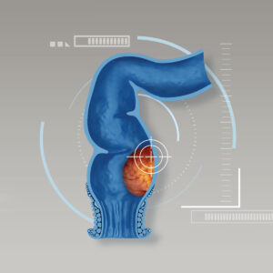
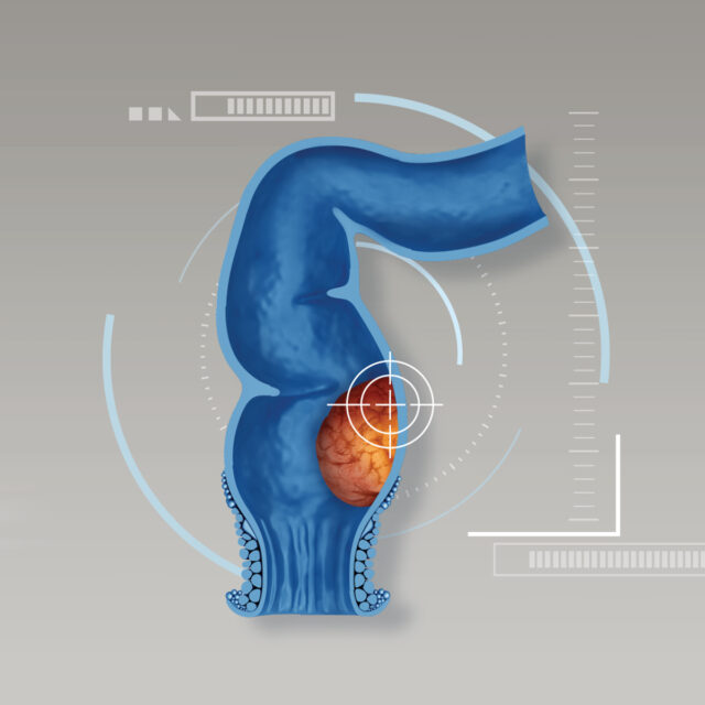
- High risk group: Family history of cancer, age over 50, blood in stool, alternating constipation and diarrhea, incomplete stool evacuation and irregular stool patterns e.g. separate hard lumps. Some symptoms are misunderstood to be only hemorrhoids. In fact, it might indicate signs of colorectal cancer.
- Treatment: If colorectal cancer is diagnosed, multidisciplinary tumor board works collaboratively with other subspecialties to design customized treatment plans collectively including radiation treatment, chemotherapy and laparoscopic surgery. Due to sphincter-saving surgical technique, bowel functions can be preserved without having a permanent colostomy. Smaller incisions and less damages to nearby areas leads to a faster recovery time with improved quality of life.
Bariatric Surgery for Treatment of Obesity
- High risk group: Being obese with BMI (body mass index) greater than 35, presented with other metabolic conditions including high blood pressure (hypertension), insulin resistance led to diabetes, dyslipidemia defined as elevated cholesterol level with reduced HDL (good lipid) as well as cardiovascular disease. Other potential risk factors also include fatty liver and obstructive sleep apnea.
- Treatment: Bariatric surgery is surgical procedures performed on the stomach or intestine to induce weight loss by the restriction of food intake and controlling hunger hormones after surgery. Not only primarily aim to reduce weight, but bariatric surgery can also dramatically reduce severity of fatty liver, improve diabetes and hypertension as well as lower risks of cardiovascular disease, resulting a better quality of life. In addition, bariatric surgery is an effective treatment for obstructive sleep apnea. Other hormone-related conditions that obtain benefits from bariatric surgery include irregular menstrual period, cysts and Polycystic ovary syndrome (PCOS).

