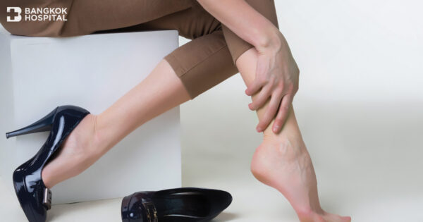
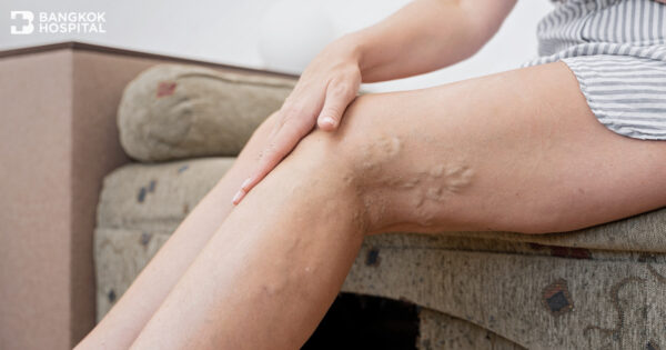
Peripheral Arterial Disease (PAD) is a common circulatory problem in which narrowed arteries reduce blood flow to the limbs e.g. the legs and arms. The severity of PAD varies, depending on the occlusive degrees, from mild discomfort to debilitating leg pain. If left untreated, this vascular disease might become chronic problem and act as a silent killer that can result in life-threatening conditions. If warning signs and symptoms of PAD exhibit, they must not be overlooked. Immediate medical attention for early detection of PAD should be sought in order to get accurate diagnosis and appropriate treatment before it progresses as well as reduce disease severity and chances of critical limb ischemia and limb amputation.
Potential risk factors of PAD
PAD is often caused by atherosclerosis, defined as deposits or atherosclerotic plaques (made up of calcium and cholesterol) build up on the artery walls and reduce blood flow. Blood vessels are comparatively similar to water supply pipes. After being used as we age, there will be plaque buildups in the pipes which limit the flood of the water. However, the accelerating factors of plaque formations include certain underlying diseases that dramatically decrease blood flow to the legs and feet, such as diabetes and chronic kidney disease. As a result, it causes painful cramping in the legs and sores on the toes, feet or legs that do not heal.
The large muscles in the calf need high amount of oxygen supply when the body requires them to function, such as walking and other exercise. At rest, the blood supply is usually sufficient. Rapid decline in oxygen and blood supply to the calf muscles while walking causes muscle cramping in calf muscles, leg numbness or weakness. If peripheral artery disease progresses with high degree of occlusion, leg pain may even occur at rest. It may be intense enough to disrupt daily activities, resulting in an impaired ambulatory function and quality of life. If disease advances, blood supply to the skin in the feet or legs drastically decreases, causing chronic wounds that do not properly heal such as diabetic foot. Critical limb ischemia can further develop when such wounds or infections progress and cause gangrene (tissue death) which amputation of the affected limb might be required.
High risk groups
PAD is commonly found in both male and female. However, people who are at higher risk for developing PAD include:
- Patients with diabetes
- Patients with hypertension
- Patients with chronic kidney disease
- Patients with dyslipidemia e.g. high blood cholesterol
- Active smokers or people who had already quit smoking. Tobacco use is a strong risk factor for developing PAD and for worsening of the disease. However, quitting smoking is associated with lower risks.
Diagnosis
Early diagnosis of PAD usually involves:
- Ankle-brachial index (ABI). ABI is a common test used to compare the blood pressure in ankle with the blood pressure in arm.
- Evaluation of vascular wall elasticity and contractility.
- Vascular ultrasound. Special ultrasound imaging techniques can help evaluate blood flow through the blood vessels and identify blocked or narrowed arteries.
- Computerized tomography (CT scan)
- Using a dye (contrast material) injected into the blood vessels, this test views blood flow through the arteries.
- Magnetic Resonance Imaging (MRI scan).
Treatments
Treatment options of PAD are depending on disease severity. Treatments include:
- Lifestyle modification. In the early stage of PAD, patients can manage the symptoms and stop disease progression through lifestyle changes including keeping underlying conditions e.g. diabetes under control, quitting smoking and having regular exercise.
- Proper foot care. Taking good care of the feet is highly advised. Diabetic patients are at risk of poor healing of sores and injuries on the lower legs and feet. Poor blood circulation can impair healing process and increase the risk of infection. Feet should be daily inspected for any abnormality or injury e.g. cuts, blisters or bruises which may not have felt happening. Annual foot check-up with a foot specialist (podiatrist) is also recommended to assess overall foot health and to identify abnormal weight bearing on the foot. Customized footwear might be needed in some patients who are prone to have sores on the foot.
- Advanced non-surgical treatment. Angioplasty is an effective option especially for the elderly or patients with underlying diseases. In this procedure, a small hollow tube called catheter is threaded to reach the affected artery. A small balloon on the tip of the catheter is inflated to reopen the narrowed artery and flatten the blockage into the artery wall, while at the same time stretching the artery to increase blood flow. A mesh framework called a stent may be inserted in the artery to help keep it open.
- Bypass surgery. If angioplasty is not applicable, bypass surgery using a vessel from another part of the body to reroute the blood supply around a blocked artery might be an alternative option.
Vascular screening
An annual vascular screening test conducted by vascular specialists substantially helps to reduce risk of developing PAD. Vascular tests usually include an ABI (ankle-brachial index) test, evaluation of vessel wall elasticity and vascular ultrasound to detect the blocked or narrowed arteries caused by atherosclerotic plaques on the vessel walls.
Diagnostic examination includes medical history review, physical examination, pulse assessment and an ABI test. An ABI test compares the blood pressure in the ankle with the blood pressure in the arm. In people who have ABI less than or equal to 0.9, it might potentially indicate blockage or occlusion in the peripheral arteries in the legs. Additional test to determine hardening arteries is further conducted by using vascular ultrasound (sound waves) to evaluate the body’s circulatory system and help identify blockages of the arteries in the legs and arms. Combined with pulse volume recording (PVR), segmental systolic blood pressure and photoplethysmography recording, analytical charts and comparisons can be obtained to determine the risk of developing PAD. The test is non-invasive and convenient with highly reliable and accurate results.
Who should have a vascular screening test
- Patients who have hardened arteries, leg pain or chronic wounds that do not heal
- Patients who have had PAD or who have family member diagnosed with PAD
- Patients with cardiovascular disease
- Smokers
- Diabetic patients
- People who have BMI greater than 30
- Patients with hypertension
- Patients with dyslipidemia
- Patients with chronic kidney disease
In addition, vascular test is also recommended in people without risk factors or any sign and symptom to screen for any abnormality that can contribute to vascular blockage in the future.
Early detection of PAD results in timely and appropriate treatment, leading to reduced disease severity and minimized chance of limb amputation. Lifestyle changes can significantly help to manage PAD-related symptoms and prevent disease progression. More importantly, to stabilize or improve PAD, these advices should be followed:
- Regular exercise of the calf and leg muscles e.g. speed walking and jogging at least 30 minutes/ day (at least 3 days/ week).
- Weight reduction, if obese.
- Eating a healthy diet. Diet low in saturated fat can help controlling blood pressure and cholesterol levels, which contribute to atherosclerosis.
- Keeping underlying diseases, including diabetes, hypertension and dyslipidemia under control.


